Trustworthy AI for breast cancer screenings (AIforScreening)
- Department Image analysis and Earth observation
- Fields involved Image analysis, Machine learning
- Industries involved Health
Breast cancer is the most prevalent form of cancer among women, and early detection is crucial for treatment and survival. In collaboration with the Cancer Registry of Norway, we are developing machine learning methods to enhance and optimise BreastScreen Norway, the national breast screening programme. This will enable a more efficient use of resources and allow healthcare professionals to dedicate more time to patients in need of further treatment.
In Norway, all women aged 50 to 69 are invited to participate in a biennial x-ray examination through the national screening programme, BreastScreen Norway. The programme is a key initiative for early breast cancer detection, helping to reduce the progression of severe disease and increase survival rates.
During the examination, images are taken of both breasts and subsequently assessed by two independent radiologists specialising in mammography. Since fewer than 1% of participants in the screening programme are diagnosed with breast cancer, a significant amount of time is spent reviewing images of healthy individuals.
Technological advancements have made it possible to automise parts of the screening process, and current research shows that machine learning models are just as accurate as human experts. Automisation frees up time and resources, making it possible for doctors to focus on patients in need of further examination and treatment.
Adaptable models for different equipment and recording conditions
We are developing methods and models for AI-based analysis of mammograms in collaboration with the Cancer Registry of Norway.
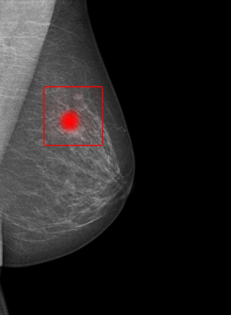
In our previous project, Machine Learning in BreastScreen Norway (MIM), we developed a prototype of a model for AI-based analysis of mammograms using a large image dataset from the Cancer Registry of Norway. During training, the model only received information about which images showed signs of cancer, but had to localise relevant areas of the breast without any further direction.
The objective of AIforScreening is not only to continue our previous research in detection, but to also examine how the models can be made more robust, as equipment and recording conditions change over time. In addition, we are studying how the contribution of time series can ensure safer identification, and how AI can be integrated in a way that facilitates productive interactions between radiologists and the models.
A two-stage method for accurate detection
It is crucial that the models aren’t so-called black boxes generating conclusions without transparency. The models should also allow for a degree of uncertainty and be able to localise possible signs of breast cancer. This is beneficial in two notable ways. Firstly, the models become more transparent and understandable. Secondly, the models themselves benefit from the explanations.
We have developed two cooperating models, where the first identifies the area of the image that most resembles cancer, and the other model zooms in and examines the area more closely. Our two-stage method has been presented at the NORA conference and our results are published in the scientific journal Nordic Machine Intelligence. Our model showed results that were on par with a radiologist, and outperformed the commercial product we compared it with.
To learn more about this project, please contact:

Project: AIforScreening: Robust and trustworthy AI for breast cancer screening
Partners: The Cancer Registry of Norway
Funding: The Research Council of Norway
Period: 2021 – 2025
Further reading:
Dahl, F. A., Brautaset, O., Holden, M., Eikvil, L., Larsen, M., & Hofvind, S. (2023). A two-stage mammography classification model using explainable-AI for ROI detection. Nordic Machine Intelligence, 3, 1-7. https://doi.org/10.5617/nmi.10459
Machine learning in BreastScreen Norway (MIM) – project page
BreastScreen Norway – kreftregisteret.no